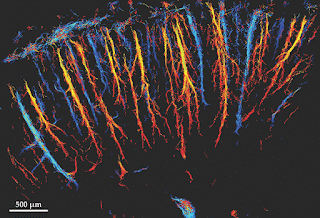
Figure 1. Superresolution ultrasound image of the blood vessels in the cortex of a rat’s brain. The colors represent velocity: Dark and light blue indicate blood flow in the direction of the skull (toward the top of the image), and red and yellow indicate flow away from the skull. (Courtesy of Mickael Tanter.)
Citation: Phys. Today 69, 2, 14 (2016); http://dx.doi.org/10.1063/PT.3.3069
Topics: Acoustic Physics, Applied Physics, Biology, Cancer, Nobel Prize, Research
With an acoustic analogue of a Nobel Prize–winning optical technique, researchers can acquire detailed images quickly.
In many ways, ultrasound waves are ideally suited to noninvasive biomedical imaging. They’re easy and inexpensive to produce and detect, and they can penetrate deep into tissue without losing their coherence or causing damage. But because of diffraction, conventional ultrasound imaging—like conventional optical microscopy—is limited in resolution to about half a wavelength. In clinical ultrasound applications, which use wavelengths between 200 µm and 1 mm, that limit precludes the imaging of many important structures, including small blood vessels. Shorter wavelengths yield better resolution, but they also penetrate less deeply into tissue.
For optical applications, innovative fluorescence techniques have been devised to overcome the diffraction limit, as recognized by the 2014 Nobel Prize in Chemistry (see Physics Today, December 2014, page 18). Inspired by that work, Mickael Tanter and his colleagues at the Langevin Institute (affiliated with ESPCI, Inserm, and CNRS) in Paris have now developed a superresolution ultrasound technique,1 which they’ve used to image the blood vessels in a rat’s brain with 10-µm resolution, as shown in figure 1. Applying the technique in humans could help to detect cancer and other diseases that alter blood-flow patterns.
Physics Today: Ultrasound resolution beats the diffraction limit, Johanna L. Miller

Comments