 |
| Source: Artificial Oxide Nanostructures: Physics of Multiscale Phenomena |
Topics: Atomic Force Microscopy, Biology, Cancer, Consumer Electronics, Electrical Engineering, Nanotechnology, Semiconductor Technology
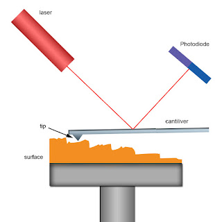
Thirty years since its first inception, the atomic force microscope has proved a hugely versatile tool. Applications range from quantifying dopant distributions in electronics and the analysis of dust particles in space, to characterizing biopsies for cancer diagnostics. More than simply bringing atomic-scale resolution to non-conducting surfaces, modifications of the technology have provided important tools for sensing chemical entities and mechanical properties, with force sensitivities so great they can be used to study and control mitosis in the proliferation of life itself. nanotechweb.org visited Basel in Switzerland, home to some of the pioneers in AFM technologies, to find out how far the field has come in the past three decades.
The development of scanning probe technologies began with the scanning tunnelling microscope (STM), and was driven by the semiconductor industry in the late 1970s. Christoph Gerber, co-inventor of the atomic force microscope, points out that although electronics feature sizes were coming close to the nanometre scale in the 1970s, there was no way of obtaining spectroscopic information of such small features. “We thought that if we established a tip very close – so that due to the proximity there would be tunnelling - we would have an instrument that could do this kind of spectroscopic work.” From there came the idea of scanning the tip and keeping the quantum tunnelling current constant. This would effectively trace a topography of the surface with a lateral resolution that could image atoms. “The big breakthrough for STM came when we were able to image the 7 × 7 reconstruction of silicon (1,1,1),” explains Gerber. As the arrangement of atoms at the surface differs from the bulk, glimpsing this reconstruction in a real image was a powerful demonstration of the instrument’s potential.
Francois Huber, who shares a lab with Hans Peter Lang, highlights how the cantilever arrays have also become useful for identifying single gene mutations from biopsies. Recently introduced cancer drugs have particularly high efficacy for specific cancer genes, such as the HER2 gene for aggressive breast cancer and the BRAF mutation found in 50% of malignant melanoma incidents. “Before we treated cancers with general chemistry or radiation – everybody got the same treatment and either you were lucky or unlucky,” says Huber. “Here we can actually target the cancer directly – it goes towards personalized medicine so that you treat patients according to their genetic predisposition.”
Nanotechweb: Atomic force microscopy – 30 years on
Comments