 |
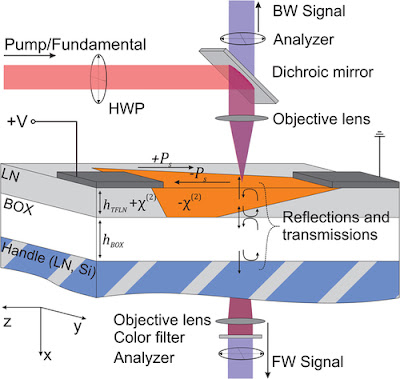
| Fig. 1 Typical geometry for the SH microscopy investigation of poled x-cut LNOI. |
Topics: Applied Physics, Optical Physics, Thin Films
Abstract
Thin film lithium niobate has been of great interest recently, and an understanding of periodically poled thin films is crucial for both fundamental physics and device developments. Second-harmonic (SH) microscopy allows for the noninvasive visualization and analysis of ferroelectric domain structures and walls. While the technique is well understood in bulk lithium niobate, SH microscopy in thin films is largely influenced by interfacial reflections and resonant enhancements, which depend on film thicknesses and substrate materials. We present a comprehensive analysis of SH microscopy in x-cut lithium niobate thin films, based on a full three-dimensional focus calculation and accounting for interface reflections. We show that the dominant signal in backreflection originates from a copropagating phase-matched process observed through reflections, rather than direct detection of the counterpropagating signal as in bulk samples. We simulate the SH signatures of domain structures by a simple model of the domain wall as an extensionless transition from a −χ(2) to a +χ(2) region. This allows us to explain the main observation of domain structures in the thin-film geometry, and, in particular, we show that the SH signal from thin poled films allows to unambiguously distinguish areas, which are completely or only partly inverted in depth.
Second harmonic microscopy of poled x-cut thin film lithium niobate: Understanding the contrast mechanism
Journal of Applied Physics 126, 114105 (2019); https://doi.org/10.1063/1.5113727
Comments